-

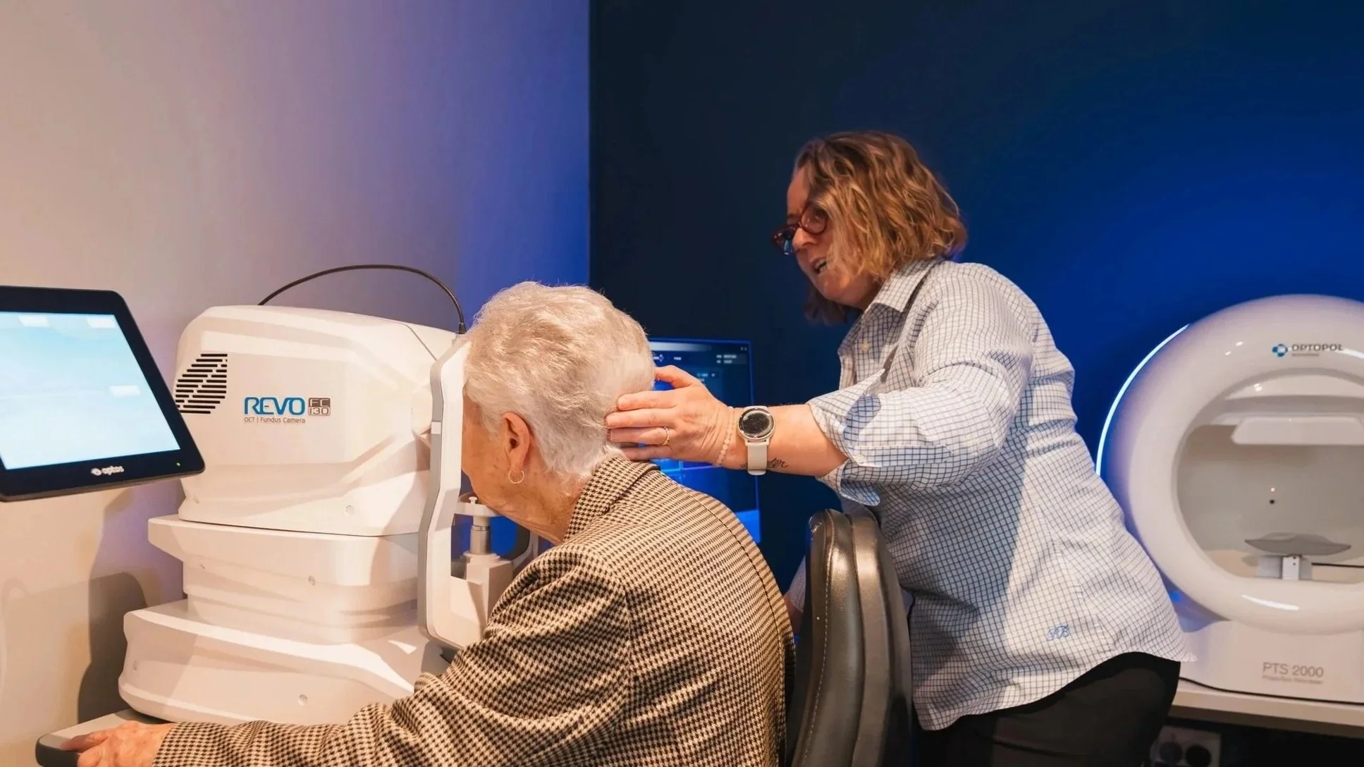
optos
The optomap ultra-widefield retinal image technology captures over 80% of your retina in one panoramic picture, while traditional methods only show about 15%. Your retina, found at the back of your eye, is the only part of your body where blood vessels can be directly observed. This allows for the detection of not only eye issues but also signs of other diseases like stroke, heart disease, high blood pressure, and diabetes. Early warning signs can appear in your retina before you notice any vision changes or feel discomfort.
The optomap exam is quick, painless, and comfortable.
-

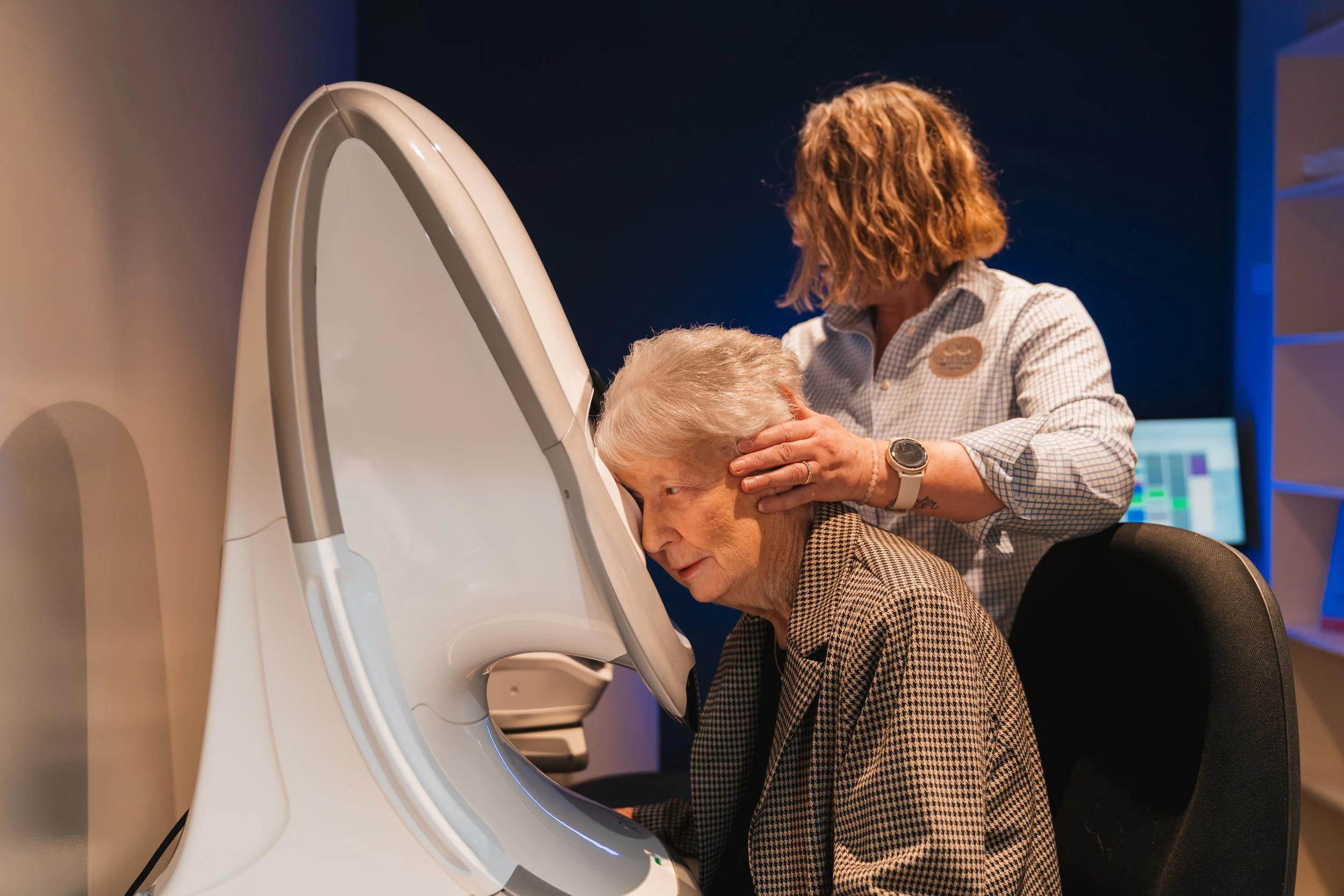
oct
OCT (Optical Coherence Tomography) is an advanced imaging test that uses light to create a cross section or layered image of the eye. The images create high resolution and 3d imaging of the retina. Our Optometrist’s are able to diagnosis ocular disease such as, macula degeneration, diabetic retinopathy and glaucoma.
This is a non-invasive test that is simply performed by resting your chin on the equipment and looking into the machine. With OCT testing all results are stored so that we are able to compare previous results to detect any ocular changes.
-

visual field
A visual field exam checks for central and side vision loss. One of our trained assistants uses a Zeiss Humphreys machine for this test. It can also find conditions like glaucoma, stroke, pituitary issues, brain tumors, and other nerve problems.
This painless test involves resting your chin on the device and looking into it with one eye, while pressing a buzzer every time you see a light flash.
This is a non-evasive exam and only takes about 20 minutes.
-

corneal topography
Corneal topography is a technique used to create a 3d map of the shape and curvature of the cornea, which is the clear front surface of the eye. It helps in diagnosing and managing various eye conditions.
The technique is performed with an instrument called a corneal topographer, which projects rings of light onto the corneas and then analyzes the reflected images using special technology.
This process is helpful for our Optometrist to diagnose, monitor and treat specific eye conditions that affect the cornea.